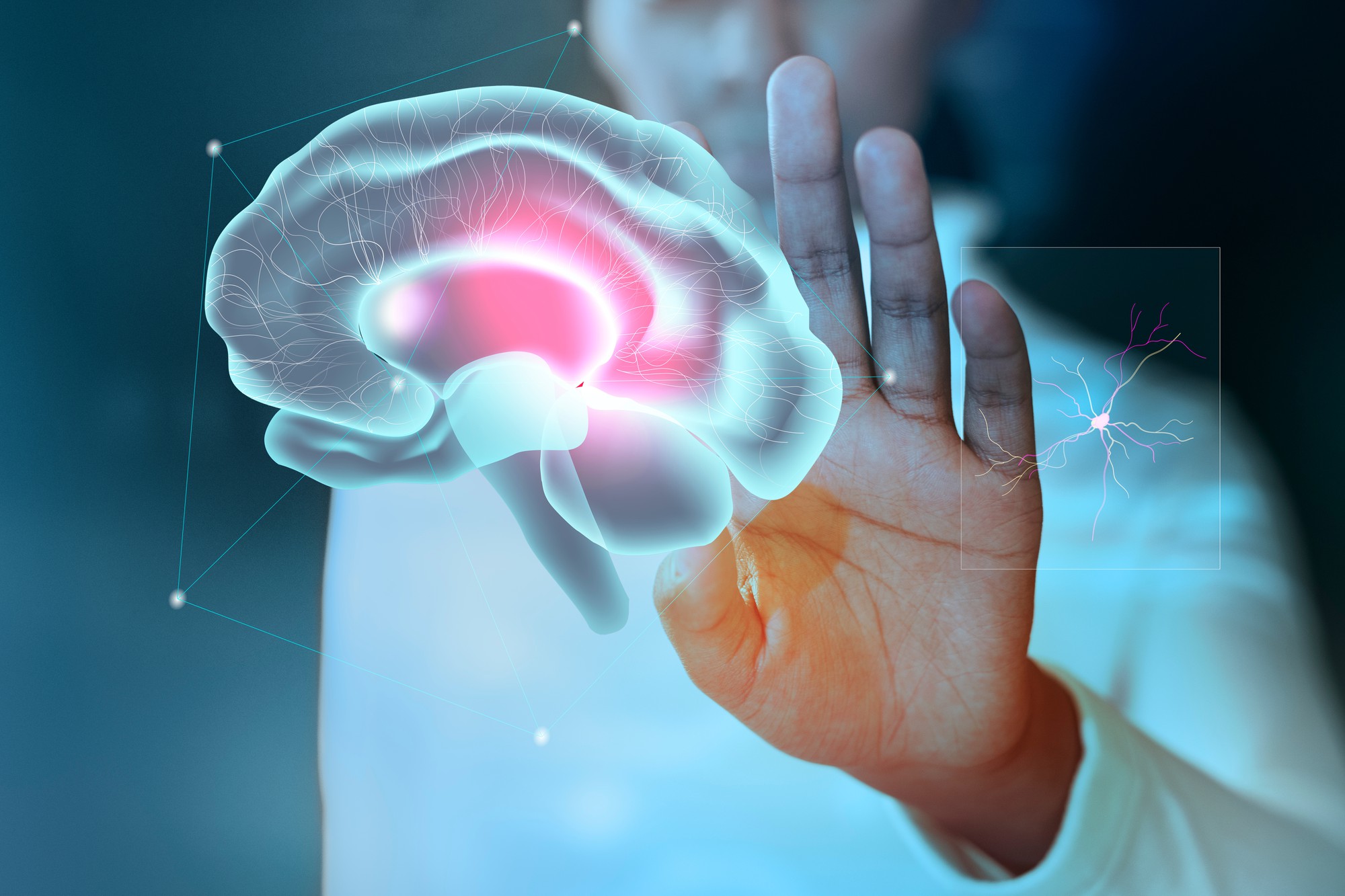

Strokes can cause irreversible, life-changing, and long-lasting brain damage. Strokes can be caused by either a blood clot, referred to as an ischemic stroke, or a burst blood vessel, referred to as a hemorrhagic stroke. Our brains require a lot of oxygen, so disruption of blood flow damages oxygen-dependent brain matter in a short period of time.
Think of the blood vessels in your brain as a perfectly engineered highway. During an ischemic stroke, a clot obstructs these vessels such that oxygen can no longer reach the brain. When an ischemic stroke occurs, immune cells known as microglia act as the “ambulances” of our body and concentrate at the site of the blood clot. Scientists divide microglia into 2 types, anti-inflammatory and pro-inflammatory. Anti-inflammatory microglia help fight brain inflammation. Pro-inflammatory microglia cause further brain inflammation by damaging nerve cells, known as neurons, and removing their protective sheath, known as myelin.
Researchers from China recently hypothesized that inhibiting an enzyme linked with inflammation, called histone deacetylase 3 or HDAC3, could decrease the production of pro-inflammatory microglia in mice. They reasoned that HDAC3 enzymes activate pro-inflammatory immune cells, so inhibiting these enzymes might alleviate inflammation.
To study how a stroke affects the production of inflammatory cells, the researchers first identified which type of cell in mice was most likely influenced by HDAC3. They induced a stroke in mice, and found that HDAC3 was more active in the mice’s microglia than in their other cells. The mice had high levels of HDAC3 in their neurons, but they weren’t active. The researchers interpreted this data to suggest that HDAC3 plays a more important role in microglia than in other types of brain cells.
Next, the researchers genetically engineered mice with no microglial HDAC3, referred to as knock-out mice. They compared the knock-out mice to a control group of mice who kept their microglial HDAC3. They induced a stroke in both groups of mice for 60 minutes. Then they used an MRI to examine the mice’s brains after 3 days, 14 days, and 35 days to see how brain damage from the stroke progressed in the 2 different groups. They found the knock-out mice had less brain damage than the control mice on all 3 days.
The researchers also found the knock-out mice scored better on movement, learning, memory, and behavioral tests than the control mice. For instance, when the scientists stuck adhesive tape to the bottom of the mice’s feet, the knock-out mice removed the tape about 50 seconds faster than their counterparts. The knock-out mice also had less myelin loss and less degeneration of electrical routes in their brains. The team interpreted these results to suggest that inhibiting microglial HDAC3 in mice prevented brain damage and increased their mental abilities after a stroke.
The researchers also tested which genes were active versus inactive in the 2 sets of mice. They found that all the pro-inflammatory genes they tested were inactive in the knock-out mice, but highly active in mice with microglial HDAC3. They also found the knock-out mice had less active genes that generated inflammatory cells.
The researchers concluded that deleting microglial HDAC3 could prevent brain inflammation, myelin removal, and brain tissue damage during a stroke. However, before HDAC3 deletion can become a therapeutic treatment they recommended future researchers determine if there are serious side effects. Decreased inflammation in mice could mean that one day the same techniques could be used to assist with decreasing the inflammatory impacts of stroke on humans.
